Various Diseases

keep an eye on your health..

Saturday, January 9, 2010

Sepsis

From Wikipedia, the free encyclopedia

Sepsis is a serious medical condition that is characterized by a whole-body inflammatory state (called a systemic inflammatory response syndrome or SIRS) and the presence of a known or suspected infection.[1][2] The body may develop this inflammatory response to microbes in the blood, urine, lungs, skin, or other tissues. An incorrect layman's term for sepsis is blood poisoning, more aptly applied to Septicemia, below.
Septicemia (also septicaemia or septicæmia [sep⋅ti⋅cæ⋅mi⋅a][3], or erroneously Septasemia and Septisema) is a related but deprecated (formerly sanctioned) medical term referring to the presence of pathogenic organisms in the blood-stream, leading to sepsis.[4] The term has not been sharply defined. It has been inconsistently used in the past by medical professionals, for example as a synonym of bacteremia, causing some confusion. The present medical consensus is therefore that the term "septicemia" is problematic and should be avoided.[2]
Sepsis is usually treated in the intensive care unit with intravenous fluids and antibiotics. If fluid replacement is insufficient to maintain blood pressure, specific vasopressor drugs can be used. Artificial ventilation and dialysis may be needed to support the function of the lungs and kidneys, respectively. To guide therapy, a central venous catheter and an arterial catheter may be placed. Sepsis patients require preventive measures for deep vein thrombosis, stress ulcers and pressure ulcers, unless other conditions prevent this. Some patients might benefit from tight control of blood sugar levels with insulin (targeting stress hyperglycemia), low-dose corticosteroids or activated drotrecogin alfa


Signs and symptoms

In addition to symptoms related to the provoking infection, sepsis is characterized by evidence of acute inflammation present throughout the entire body, and is, therefore, frequently associated with fever and elevated white blood cell count (leukocytosis) or low white blood cell count and lower-than-average temperature, and vomiting[citation needed]. The modern concept of sepsis is that the host's immune response to the infection causes most of the symptoms of sepsis, resulting in hemodynamic consequences and damage to organs. This host response has been termed systemic inflammatory response syndrome (SIRS) and is characterized by hemodynamic compromise and resultant metabolic derangement. Outward physical symptoms of this response frequently include a high heart rate (above 100 beats per minute), high respiratory rate (above 20 breaths per minute), elevated WBC count (above 12,000) and elevated or lowered body temperature (under 36 °C or over 38 °C). Sepsis is differentiated from SIRS by the presence of a known pathogen. For example SIRS and a positive blood culture for a pathogen indicates the presence of sepsis. Without a known infection you can not classify the above symptoms as sepsis, only SIRS.
This immunological response causes widespread activation of acute-phase proteins, affecting the complement system and the coagulation pathways, which then cause damage to the vasculature as well as to the organs. Various neuroendocrine counter-regulatory systems are then activated as well, often compounding the problem. Even with immediate and aggressive treatment, this may progress to multiple organ dysfunction syndrome and eventually death.


Definition

According to the American College of Chest Physicians and the Society of Critical Care Medicine[2], there are different levels of sepsis:
Examples of end-organ dysfunction include the following:[10]
More specific definitions of end-organ dysfunction exist for SIRS in pediatrics.[11]
  • Cardiovascular dysfunction (after fluid resuscitation with at least 40 ml/kg of crystalloid)
    • hypotension with blood pressure < 5th percentile for age or systolic blood pressure < 2 standard deviations below normal for age, OR
    • vasopressor requirement, OR
    • two of the following criteria:
  • Respiratory dysfunction (in the absence of cyanotic heart disease or known chronic lung disease)
    • the ratio of the arterial partial-pressure of oxygen to the fraction of oxygen in the gases inspired (PaO2/FiO2) < 300 (the definition of acute lung injury), OR
    • arterial partial-pressure of carbon dioxide (PaCO2) > 65 torr (20 mmHg) over baseline PaCO2 (evidence of hypercapnic respiratory failure), OR
    • supplemental oxygen requirement of greater than FiO2 0.5 to maintain oxygen saturation ≥ 92%
  • Neurologic dysfunction
  • Hematologic dysfunction
  • Renal dysfunction
  • Hepatic dysfunction (only applicable to infants > 1 month)
Consensus definitions, however, continue to evolve, with the latest expanding the list of signs and symptoms of sepsis to reflect clinical bedside experience

Treatment

Adults and children

The therapy of sepsis rests on antibiotics, surgical drainage of infected fluid collections, fluid replacement and appropriate support for organ dysfunction. This may include hemodialysis in kidney failure, mechanical ventilation in pulmonary dysfunction, transfusion of blood products, and drug and fluid therapy for circulatory failure. Ensuring adequate nutrition—preferably by enteral feeding, but if necessary by parenteral nutrition—is important during prolonged illness.
A problem in the adequate management of septic patients has been the delay in administering therapy after sepsis has been recognized. Published studies have demonstrated that for every hour delay in the administration of appropriate antibiotic therapy there is an associated 7% rise in mortality. A large international collaboration was established to educate people about sepsis and to improve patient outcomes with sepsis, entitled the "Surviving Sepsis Campaign." The Campaign has published an evidence-based review of management strategies for severe sepsis,[5] with the aim to publish a complete set of guidelines in subsequent years.
Early Goal Directed Therapy (EGDT), developed at Henry Ford Hospital by E. Rivers, MD, is a systematic approach to resuscitation that has been validated in the treatment of severe sepsis and septic shock. It is meant to be started in the Emergency Department. The theory is that one should use a step-wise approach, having the patient meet physiologic goals, to optimize cardiac preload, afterload, and contractility, thus optimizing oxygen delivery to the tissues.[13] A recent meta-analysis showed that EGDT provides a benefit on mortality in patients with sepsis.[14] As of December 2008 some controversy around its uses remains and a number of trials are ongoing in an attempt to resolve this.[15]
In EGDT, fluids are administered until the central venous pressure (CVP), as measured by a central venous catheter, reaches 8-12 cm of water (or 10-15 cm of water in mechanically ventilated patients). Rapid administration of several liters of isotonic crystalloid solution is usually required to achieve this. If the mean arterial pressure is less than 65 mmHg or greater than 90 mmHg, vasopressors or vasodilators are given as needed to reach the goal. Once these goals are met, the mixed venous oxygen saturation (SvO2), i.e., the oxygen saturation of venous blood as it returns to the heart as measured at the vena cava, is optimized. If the SvO2 is less than 70%, blood is given to reach a hemoglobin of 10 g/dl and then inotropes are added until the SvO2 is optimized. Elective intubation may be performed to reduce oxygen demand if the SvO2 remains low despite optimization of hemodynamics. Urine output is also monitored, with a minimum goal of 0.5 ml/kg/h. In the original trial, mortality was cut from 46.5% in the control group to 30.5% in the intervention group.[13] The Surviving Sepsis Campaign guidelines recommend EGDT for the initial resuscitation of the septic patient with a level B strength of evidence (single randomized control trial).[5]
Most therapies aimed at the inflammation process itself have failed to improve outcome, however drotrecogin alfa (activated protein C, one of the coagulation factors) has been shown to decrease mortality from about 31% to about 25% in severe sepsis. To qualify for drotrecogin alfa, a patient must have severe sepsis or septic shock with an APACHE II score of 25 or greater and a low risk of bleeding.[16]
During critical illness, a state of adrenal insufficiency and tissue resistance (the word 'relative' resistance should be avoided[17]) to corticosteroids may occur. This has been termed critical illness–related corticosteroid insufficiency.[17] Treatment with corticosteroids might be most beneficial in those with septic shock and early severe acute respiratory distress syndrome (ARDS), whereas its role in other patients such as those with pancreatitis or severe pneumonia is unclear.[17] These recommendations stem from studies showing benefits from low dose hydrocortisone treatment for septic shock patients and methylprednisolone in ARDS patients.[18][19][20][21][22][23] However, the exact way of determining corticosteroid insufficiency remains problematic. It should be suspected in those poorly responding to resuscitation with fluids and vasopressors. ACTH stimulation testing is not recommended to confirm the diagnosis.[17] Glucocorticoid drugs should be weaned and not stopped abruptly.
In some cases, sepsis may lead to inadequate tissue perfusion and necrosis. As this may affect the extremities, amputation may become necessary. On January 8 2009 a patent request was submitted for the possible treatment of sepsis ( 20090011974 Scavenger Receptor B1 (Cla-1) Targeting for the Treatment of Infection, Sepsis and Inflammation 01-08-2009) .


Pneumonia

From Wikipedia, the free encyclopedia

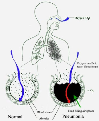
Pneumonia is an abnormal inflammatory condition of the lung.[1] It is often characterized as including inflammation of the parenchyma of the lung (that is, the alveoli) and abnormal alveolar filling with fluid (consolidation and exudation).[2]
The alveoli are microscopic air-filled sacs in the lungs responsible for absorbing oxygen. Pneumonia can result from a variety of causes, including infection with bacteria, viruses, fungi, or parasites, and chemical or physical injury to the lungs. Its cause may also be officially described as idiopathic—that is, unknown—when infectious causes have been excluded.
Typical symptoms associated with pneumonia include cough, chest pain, fever, and difficulty in breathing. Diagnostic tools include x-rays and examination of the sputum. Treatment depends on the cause of pneumonia; bacterial pneumonia is treated with antibiotics.
Pneumonia is a common illness which occurs in all age groups, and is a leading cause of death among the elderly and people who are chronically and terminally ill. Additionally, it is the leading cause of death in children under five years old worldwide.[3] Vaccines to prevent certain types of pneumonia are available. The prognosis depends on the type of pneumonia, the appropriate treatment, any complications, and the person's underlying health.


Classification

Pneumonias can be classified in several ways. Pathologists originally classified them according to the anatomic changes that were found in the lungs during autopsies. As more became known about the microorganisms causing pneumonia, a microbiologic classification arose, and with the advent of x-rays, a radiological classification. Another important system of classification is the combined clinical classification, which combines factors such as age, risk factors for certain microorganisms, the presence of underlying lung disease and underlying systemic disease, and whether the person has recently been hospitalized.

Other types of pneumonia

SARS is a highly contagious and deadly type of pneumonia which first occurred in 2002 after initial outbreaks in China. SARS is caused by the SARS coronavirus, a previously unknown pathogen.
BOOP is caused by inflammation of the small airways of the lungs. It is also known as cryptogenic organizing pneumonitis (COP).
Eosinophilic pneumonia is invasion of the lung by eosinophils, a particular kind of white blood cell. Eosinophilic pneumonia often occurs in response to infection with a parasite or after exposure to certain types of environmental factors.
Chemical pneumonia (usually called chemical pneumonitis) is caused by chemical toxicants such as pesticides, which may enter the body by inhalation or by skin contact. When the toxic substance is an oil, the pneumonia may be called lipoid pneumonia.
Aspiration pneumonia (or aspiration pneumonitis) is caused by aspirating foreign objects which are usually oral or gastric contents, either while eating, or after reflux or vomiting which results in bronchopneumonia. The resulting lung inflammation is not an infection but can contribute to one, since the material aspirated may contain anaerobic bacteria or other unusual causes of pneumonia. Aspiration is a leading cause of death among hospital and nursing home patients, since they often cannot adequately protect their airways and may have otherwise impaired defenses.
Dust pneumonia describes disorders caused by excessive exposure to dust storms, particularly during the Dust Bowl in the United States. With dust pneumonia, dust settles all the way into the alveoli of the lungs, stopping the cilia from moving and preventing the lungs from ever clearing themselves.

Signs and symptoms

People with infectious pneumonia often have a cough producing greenish or yellow sputum, or phlegm and a high fever that may be accompanied by shaking chills. Shortness of breath is also common, as is pleuritic chest pain, a sharp or stabbing pain, either experienced during deep breaths or coughs or worsened by them. People with pneumonia may cough up blood, experience headaches, or develop sweaty and clammy skin. Other possible symptoms are loss of appetite, fatigue, blueness of the skin, nausea, vomiting, mood swings, and joint pains or muscle aches. Less common forms of pneumonia can cause other symptoms; for instance, pneumonia caused by Legionella may cause abdominal pain and diarrhea, while pneumonia caused by tuberculosis or Pneumocystis may cause only weight loss and night sweats. In elderly people manifestations of pneumonia may not be typical. They may develop a new or worsening confusion or may experience unsteadiness, leading to falls. Infants with pneumonia may have many of the symptoms above, but in many cases they are simply sleepy or have a decreased appetite.


Symptoms of pneumonia need immediate medical evaluation. Physical examination by a health care provider may reveal fever or sometimes low body temperature, an increased respiratory rate, low blood pressure, a high heart rate, or a low oxygen saturation, which is the amount of oxygen in the blood as indicated by either pulse oximetry or blood gas analysis. People who are struggling to breathe, who are confused, or who have cyanosis (blue-tinged skin) require immediate attention.
Physical examination of the lungs may be normal, but often shows decreased expansion of the chest on the affected side, bronchial breathing on auscultation with a stethoscope (harsher sounds from the larger airways transmitted through the inflamed and consolidated lung), and rales (or crackles) heard over the affected area during inspiration. Percussion may be dulled over the affected lung, but increased rather than decreased vocal resonance (which distinguishes it from a pleural effusion).[8] While these signs are relevant, they are insufficient to diagnose or rule out a pneumonia; moreover, in studies it has been shown that two doctors can arrive at different findings on the same patient.


Cause

Pneumonia can be caused by microorganisms, irritants and unknown causes. When pneumonias are grouped this way, infectious causes are the most common type.
The symptoms of infectious pneumonia are caused by the invasion of the lungs by microorganisms and by the immune system's response to the infection. Although more than one hundred strains of microorganism can cause pneumonia, only a few are responsible for most cases. The most common causes of pneumonia are viruses and bacteria. Less common causes of infectious pneumonia are fungi and parasites.

Diagnosis

If pneumonia is suspected on the basis of a patient's symptoms and findings from physical examination, further investigations are needed to confirm the diagnosis. Information from a chest X-ray and blood tests are helpful, and sputum cultures in some cases. The chest X-ray is typically used for diagnosis in hospitals and some clinics with X-ray facilities. However, in a community setting (general practice), pneumonia is usually diagnosed based on symptoms and physical examination alone.[citation needed] Diagnosing pneumonia can be difficult in some people, especially those who have other illnesses. Occasionally a chest CT scan or other tests may be needed to distinguish pneumonia from other illnesses.

Investigations

An important test for pneumonia in unclear situations is a chest x-ray. Chest x-rays can reveal areas of opacity (seen as white) which represent consolidation. Pneumonia is not always seen on x-rays, either because the disease is only in its initial stages, or because it involves a part of the lung not easily seen by x-ray. In some cases, chest CT (computed tomography) can reveal pneumonia that is not seen on chest x-ray. X-rays can be misleading, because other problems, like lung scarring and congestive heart failure, can mimic pneumonia on x-ray.[12] Chest x-rays are also used to evaluate for complications of pneumonia (see below.)
If antibiotics fail to improve the patient's health, or if the health care provider has concerns about the diagnosis, a culture of the person's sputum may be requested. Sputum cultures generally take at least two to three days, so they are mainly used to confirm that the infection is sensitive to an antibiotic that has already been started. A blood sample may similarly be cultured to look for bacteria in the blood. Any bacteria identified are then tested to see which antibiotics will be most effective.
A complete blood count may show a high white blood cell count, indicating the presence of an infection or inflammation. In some people with immune system problems, the white blood cell count may appear deceptively normal. Blood tests may be used to evaluate kidney function (important when prescribing certain antibiotics) or to look for low blood sodium. Low blood sodium in pneumonia is thought to be due to extra anti-diuretic hormone produced when the lungs are diseased (SIADH). Specific blood serology tests for other bacteria (Mycoplasma, Legionella and Chlamydophila) and a urine test for Legionella antigen are available. Respiratory secretions can also be tested for the presence of viruses such as influenza, respiratory syncytial virus, and adenovirus. Liver function tests should be carried out to test for damage caused by sepsis.


Prevention

There are several ways to prevent infectious pneumonia. Appropriately treating underlying illnesses (such as AIDS) can decrease a person's risk of pneumonia. Smoking cessation is important not only because it helps to limit lung damage, but also because cigarette smoke interferes with many of the body's natural defenses against pneumonia.
Research shows that there are several ways to prevent pneumonia in newborn infants. Testing pregnant women for Group B Streptococcus and Chlamydia trachomatis, and then giving antibiotic treatment if needed, reduces pneumonia in infants. Suctioning the mouth and throat of infants with meconium-stained amniotic fluid decreases the rate of aspiration pneumonia.
Vaccination is important for preventing pneumonia in both children and adults. Vaccinations against Haemophilus influenzae and Streptococcus pneumoniae in the first year of life have greatly reduced the role these bacteria play in causing pneumonia in children. Vaccinating children against Streptococcus pneumoniae has also led to a decreased incidence of these infections in adults because many adults acquire infections from children. Hib vaccine is now widely used around the globe. The childhood pneumococcal vaccine is still as of 2009 predominantly used in high-income countries, though this is changing. In 2009, Rwanda became the first low-income country to introduce pneumococcal conjugate vaccine into their national immunization program.[16]
A vaccine against Streptococcus pneumoniae is also available for adults. In the U.S., it is currently recommended for all healthy individuals older than 65 and any adults with emphysema, congestive heart failure, diabetes mellitus, cirrhosis of the liver, alcoholism, cerebrospinal fluid leaks, or those who do not have a spleen. A repeat vaccination may also be required after five or ten years.[17]
Influenza vaccines should be given yearly to the same individuals who receive vaccination against Streptococcus pneumoniae. In addition, health care workers, nursing home residents, and pregnant women should receive the vaccine.[18] When an influenza outbreak is occurring, medications such as amantadine, rimantadine, zanamivir, and oseltamivir can help prevent influenza.

Treatment

Most cases of pneumonia can be treated without hospitalization. Typically, oral antibiotics, rest, fluids, and home care are sufficient for complete resolution. However, people with pneumonia who are having trouble breathing, people with other medical problems, and the elderly may need more advanced treatment. If the symptoms get worse, the pneumonia does not improve with home treatment, or complications occur, the person will often have to be hospitalized.

Bacterial pneumonia

Antibiotics are used to treat bacterial pneumonia. In contrast, antibiotics are not useful for viral pneumonia, although they sometimes are used to treat or prevent bacterial infections that can occur in lungs damaged by a viral pneumonia.[citation needed] The antibiotic choice depends on the nature of the pneumonia, the most common microorganisms causing pneumonia in the local geographic area, and the immune status and underlying health of the individual. Treatment for pneumonia should ideally be based on the causative microorganism and its known antibiotic sensitivity. However, a specific cause for pneumonia is identified in only 50% of people, even after extensive evaluation.[citation needed] Because treatment should generally not be delayed in any person with a serious pneumonia, empiric treatment is usually started well before laboratory reports are available. In the United Kingdom, amoxicillin and clarithromycin or erythromycin are the antibiotics selected for most patients with community-acquired pneumonia; patients allergic to penicillins are given erythromycin instead of amoxicillin.[21] In North America, where the "atypical" forms of community-acquired pneumonia are becoming more common, macrolides (such as azithromycin and clarithromycin), the fluoroquinolones, and doxycycline have displaced amoxicillin as first-line outpatient treatment for community-acquired pneumonia.[22] The duration of treatment has traditionally been seven to ten days, but there is increasing evidence that shorter courses (as short as three days) are sufficient.[23][24][25]
Antibiotics for hospital-acquired pneumonia include third- and fourth-generation cephalosporins, carbapenems, fluoroquinolones, aminoglycosides, and vancomycin.[26] These antibiotics are usually given intravenously. Multiple antibiotics may be administered in combination in an attempt to treat all of the possible causative microorganisms. Antibiotic choices vary from hospital to hospital because of regional differences in the most likely microorganisms, and because of differences in the microorganisms' abilities to resist various antibiotic treatments.
People who have difficulty breathing due to pneumonia may require extra oxygen. Extremely sick individuals may require intensive care, often including endotracheal intubation and artificial ventilation.
Over the counter cough medicine has not been found to be helpful in pneumonia.[27]

Viral pneumonia

Viral pneumonia caused by influenza A may be treated with rimantadine or amantadine, while viral pneumonia caused by influenza A or B may be treated with oseltamivir or zanamivir. These treatments are beneficial only if they are started within 48 hours of the onset of symptoms. Many strains of H5N1 influenza A, also known as avian influenza or "bird flu," have shown resistance to rimantadine and amantadine. There are no known effective treatments for viral pneumonias caused by the SARS coronavirus, adenovirus, hantavirus, or parainfluenza virus.

Aspiration pneumonia

There is no evidence to support the use of antibiotics in chemical pneumonitis without bacterial infection. If infection is present in aspiration pneumonia, the choice of antibiotic will depend on several factors, including the suspected causative organism and whether pneumonia was acquired in the community or developed in a hospital setting. Common options include clindamycin, a combination of a beta-lactam antibiotic and metronidazole, or an aminoglycoside.[28] Corticosteroids are commonly used in aspiration pneumonia, but there is no evidence to support their use either


Cholera

From Wikipedia, the free encyclopedia


Cholera, sometimes known as Asiatic or epidemic cholera, is an infectious gastroenteritis caused by enterotoxin-producing strains of the bacterium Vibrio cholerae.[1][2] Transmission to humans occurs through eating food or drinking water contaminated with Vibrio cholerae from other cholera patients. The major reservoir for cholera was long assumed to be humans themselves, but considerable evidence exists that aquatic environments can serve as reservoirs of the bacteria.
Vibrio cholerae is a Gram-negative bacterium that produces cholera toxin, an enterotoxin, whose action on the mucosal epithelium lining of the small intestine is responsible for the disease's most salient characteristic, exhaustive diarrhea.[1] In its most severe forms, cholera is one of the most rapidly fatal illnesses known, and a healthy person's blood pressure may drop to hypotensive levels within an hour of the onset of symptoms; infected patients may die within three hours if medical treatment is not provided.[1] In a common scenario, the disease progresses from the first liquid stool to shock in 4 to 12 hours, with death following in 18 hours to several days, unless oral (or, in more serious cases, intravenous) rehydration therapy is provided.[3][4]
It is estimated that most cases of cholera are unreported due to poor surveillance systems, particularly in Africa. Fatality rates are 5% of total cases in Africa, and less than 1% elsewhere.[5] For a map of recent international outbreaks, see:[3]


Treatment

In most cases cholera can be successfully treated with oral rehydration therapy. Prompt replacement of water and electrolytes is the principal treatment for cholera, as dehydration and electrolyte depletion occur rapidly. Oral rehydration therapy or ORT is highly effective, safe, and simple to administer. In situations where commercially produced ORT sachets are too expensive or difficult to obtain, alternative homemade solutions using various formulas of water, sugar, table salt, baking soda, and fruit offer less expensive methods of electrolyte repletion. In severe cholera cases with significant dehydration and the administration of intravenous rehydration solutions may be necessary.
Antibiotics shorten the course of the disease, and reduce the severity of the symptoms. However oral rehydration therapy remains the principal treatment. Tetracycline is typically used as the primary antibiotic, although some strains of V. cholerae exist that have shown resistance. Other antibiotics that have been proven effective against V. cholerae include cotrimoxazole, erythromycin, doxycycline, chloramphenicol, and furazolidone.[6] Fluoroquinolones such as norfloxacin also may be used, but resistance has been reported.[7]
Rapid diagnostic assay methods are available for the identification of multidrug resistant V. cholerae.[8] New generation antimicrobials have been discovered which are effective against V. cholerae in in vitro studies.[9]
The success of treatment is significantly affected by the speed and method of treatment. If cholera patients are treated quickly and properly, the mortality rate is less than 1%; however, with untreated cholera the mortality rate rises to 50–60%.

Diagnosis

In epidemic situations a clinical diagnosis is made by taking a history of symptoms from the patient and by a brief examination only. Treatment is usually started without or before confirmation by laboratory analysis of specimens.
Stool and swab samples collected in the acute stage of the disease, before antibiotics have been administered, are the most useful specimens for laboratory diagnosis. If an epidemic of cholera is suspected, the most common causative agent is Vibrio cholerae O1. If V. cholerae serogroup O1 is not isolated, the laboratory should test for V. cholerae O139. However, if neither of these organisms is isolated, it is necessary to send stool specimens to a reference laboratory. Infection with V. cholerae O139 should be reported and handled in the same manner as that caused by V. cholerae O1. The associated diarrheal illness should be referred to as cholera and must be reported as a case of cholera to the appropriate public health authorities.[15]
A number of special media have been employed for the cultivation for cholera vibrios. They are classified as follows:

Holding or transport media

  1. Venkataraman-Ramakrishnan (VR) medium: This medium has 20g Sea Salt Powder and 5g Peptone dissolved in 1L of distilled water.
  2. Cary-Blair medium: This is the most widely-used carrying medium. This is a buffered solution of sodium chloride, sodium thioglycollate, disodium phosphate and calcium chloride at pH 8.4.
  3. Autoclaved sea water

Enrichment media

  1. Alkaline peptone water at pH 8.6
  2. Monsur's taurocholate tellurite peptone water at pH 9.2

Plating media

  1. Alkaline bile salt agar (BSA): The colonies are very similar to those on nutrient agar.
  2. Monsur's gelatin Tauro cholate trypticase tellurite agar (GTTA) medium: Cholera vibrios produce small translucent colonies with a greyish black centre.
  3. TCBS medium: This the mostly widely used medium. This medium contains thiosulphate, citrate, bile salts and sucrose. Cholera vibrios produce flat 2–3 mm in diameter, yellow nucleated colonies.
Direct microscopy of stool is not recommended as it is unreliable. Microscopy is preferred only after enrichment, as this process reveals the characteristic motility of Vibrios and its inhibition by appropriate antiserum. Diagnosis can be confirmed as well as serotyping done by agglutination with specific sera.

Biochemistry

Most of the V. cholerae bacteria in the contaminated water consumed by the host do not survive the highly acidic conditions of the human stomach.[24] The few bacteria that do survive conserve their energy and stored nutrients during the passage through the stomach by shutting down much protein production. When the surviving bacteria exit the stomach and reach the small intestine, they need to propel themselves through the thick mucus that lines the small intestine to get to the intestinal wall where they can thrive. V. cholerae bacteria start up production of the hollow cylindrical protein flagellin to make flagella, the curly whip-like tails that they rotate to propel themselves through the mucus that lines the small intestine.
Once the cholera bacteria reach the intestinal wall, they do not need the flagella propellers to move themselves any longer. The bacteria stop producing the protein flagellin, thus again conserving energy and nutrients by changing the mix of proteins that they manufacture in response to the changed chemical surroundings. On reaching the intestinal wall, V. cholerae start producing the toxic proteins that give the infected person a watery diarrhea. This carries the multiplying new generations of V. cholerae bacteria out into the drinking water of the next host—if proper sanitation measures are not in place.


Microbiologists have studied the genetic mechanisms by which the V. cholerae bacteria turn off the production of some proteins and turn on the production of other proteins as they respond to the series of chemical environments they encounter, passing through the stomach, through the mucous layer of the small intestine, and on to the intestinal wall.[25] Of particular interest have been the genetic mechanisms by which cholera bacteria turn on the protein production of the toxins that interact with host cell mechanisms to pump chloride ions into the small intestine, creating an ionic pressure which prevents sodium ions from entering the cell. The chloride and sodium ions create a salt water environment in the small intestines which through osmosis can pull up to six liters of water per day through the intestinal cells creating the massive amounts of diarrhea. The host can become rapidly dehydrated if an appropriate mixture of dilute salt water and sugar is not taken to replace the blood's water and salts lost in the diarrhea.
By inserting separate, successive sections of V. cholerae DNA into the DNA of other bacteria such as E. coli that would not naturally produce the protein toxins, researchers have investigated the mechanisms by which V. cholerae responds to the changing chemical environments of the stomach, mucous layers, and intestinal wall. Researchers have discovered that there is a complex cascade of regulatory proteins that control expression of V. cholerae virulence determinants. In responding to the chemical environment at the intestinal wall, the V. cholerae bacteria produce the TcpP/TcpH proteins, which, together with the ToxR/ToxS proteins, activate the expression of the ToxT regulatory protein. ToxT then directly activates expression of virulence genes that produce the toxins that cause diarrhea in the infected person and that permit the bacteria to colonize the intestine.[25] Current research aims at discovering "the signal that makes the cholera bacteria stop swimming and start to colonize (that is, adhere to the cells of) the small intestine."


Dengue fever

From Wikipedia, the free encyclopedia

Dengue fever (pronounced UK: /ˈdɛŋɡeɪ/, US: /ˈdɛŋɡiː/) and dengue hemorrhagic fever (DHF) are acute febrile diseases which occur in the tropics, can be life-threatening, and are caused by four closely related virus serotypes of the genus Flavivirus, family Flaviviridae.[1] It is also known as breakbone fever. It occurs widely in the tropics, including northern Argentina, northern Australia, the entirety of Bangladesh, Barbados, Bolivia[2], Brazil, Cambodia, Costa Rica, Dominican Republic, Guatemala, Guyana, Honduras, India, Indonesia, Jamaica, Laos, Malaysia, Mexico, Pakistan, Panama, Paraguay[3], Philippines, Puerto Rico, Samoa[4], Singapore, Sri Lanka, Suriname, Taiwan, Thailand, Trinidad, Venezuela and Vietnam, and increasingly in southern China[5]. Unlike malaria, dengue is just as prevalent in the urban districts of its range as in rural areas. Each serotype is sufficiently different that there is no cross-protection and epidemics caused by multiple serotypes (hyperendemicity) can occur. Dengue is transmitted to humans by the Aedes aegypti or more rarely the Aedes albopictus mosquito, which feed during the day.[6]
The WHO says some 2.5 billion people, two fifths of the world's population, are now at risk from dengue and estimates that there may be 50 million cases of dengue infection worldwide every year. The disease is now endemic in more than 100 countries.[7]


Signs and symptoms

The disease manifests as a sudden onset of severe headache, muscle and joint pains (myalgias and arthralgias—severe pain that gives it the nickname break-bone fever or bonecrusher disease), fever, and rash.[8] The dengue rash is characteristically bright red petechiae and usually appears first on the lower limbs and the chest; in some patients, it spreads to cover most of the body. There may also be gastritis with some combination of associated abdominal pain, nausea, vomiting, or diarrhea.
Some cases develop much milder symptoms which can be misdiagnosed as influenza or other viral infection when no rash is present. Thus travelers from tropical areas may pass on dengue inadvertently, having not been properly diagnosed at the height of their illness. Patients with dengue can pass on the infection only through mosquitoes or blood products and only while they are still febrile. The classic dengue fever lasts about two to seven days, with a smaller peak of fever at the trailing end of the disease (the so-called "biphasic pattern"). Clinically, the platelet count will drop until the patient's temperature is normal. Cases of DHF also show higher fever, variable hemorrhagic phenomena, thrombocytopenia, and hemoconcentration. A small proportion of cases lead to dengue shock syndrome (DSS) which has a high mortality rate. DHF combined with a cirrhotic liver has been suspected in rapid development of hepatocellular carcinoma (HCC). Given that the dengue virus (DEN) is related to the Hepatitis C virus, this is an avenue for further research as HCC is among the top five cancerous causes of death outside Europe and North America. Normally HCC does not occur in a cirrhotic liver for ten or more years after the cessation of the poisoning agent. DHF patients can develop HCC within one year of cessation of poisoning agent.

Diagnosis

The diagnosis of dengue is usually made clinically. The classic picture is high fever with no localising source of infection, a petechial rash with thrombocytopenia and relative leukopenia - low platelet and white blood cell count. Care has to be taken as diagnosis of DHF can mask end stage liver disease and vice versa.
  1. Fever, bladder problem, constant headaches, eye pain, severe dizziness and loss of appetite.
  2. Hemorrhagic tendency (positive tourniquet test, spontaneous bruising, bleeding from mucosa, gingiva, injection sites, etc.; vomiting blood, or bloody diarrhea)
  3. Thrombocytopenia (<100,000 platelets per mm³ or estimated as less than 3 platelets per high power field)
  4. Evidence of plasma leakage (hematocrit more than 20% higher than expected, or drop in hematocrit of 20% or more from baseline following IV fluid, pleural effusion, ascites, hypoproteinemia)
  5. Encephalitic occurrences.
Dengue shock syndrome is defined as dengue hemorrhagic fever plus:
  • Weak rapid pulse,
  • Narrow pulse pressure (less than 20 mm Hg)
  • Cold, clammy skin and restlessness.
Dependable, immediate diagnosis of dengue can be performed in rural areas by the use of Rapid Diagnostic Test kits, which also differentiate between primary and secondary dengue infections.[9] Serology and polymerase chain reaction (PCR) studies are available to confirm the diagnosis of dengue if clinically indicated. Dengue can be a life threatening fever.

Prevention

Vaccine development

There is no tested and approved vaccine for the dengue flavivirus. There are many ongoing vaccine development programs. Among them is the Pediatric Dengue Vaccine Initiative set up in 2003 with the aim of accelerating the development and introduction of dengue vaccine(s) that are affordable and accessible to poor children in endemic countries.[11] Thai researchers are testing a dengue fever vaccine on 3,000–5,000 human volunteers after having successfully conducted tests on animals and a small group of human volunteers.[12] A number of other vaccine candidates are entering phase I or II testing.[13]

Mosquito control

Primary prevention of dengue mainly resides in mosquito control. There are two primary methods: larval control and adult mosquito control.[citation needed] In urban areas, Aedes mosquitos breed on water collections in artificial containers such as plastic cups, used tires, broken bottles, flower pots, etc. Periodic draining or removal of containers is the most effective way of reducing the breeding grounds for mosquitos.[citation needed] Larvicide treatment is another effective way to control the vector larvae, but the larvicide chosen should be long-lasting and preferably have World Health Organization clearance for use in drinking water. There are some very effective insect growth regulators (IGRs) available which are both safe and long-lasting (e.g., pyriproxyfen). For reducing the adult mosquito load, fogging with insecticide is somewhat effective.[citation needed]
Prevention of mosquito bites is another way of preventing disease. This can be achieved by using insect repellent, mosquito traps or mosquito nets.

Treatment

The mainstay of treatment is timely supportive therapy to tackle shock due to hemoconcentration and bleeding. Close monitoring of vital signs in critical period (between day 2 to day 7 of fever) is critical. Increased oral fluid intake is recommended to prevent dehydration. Supplementation with intravenous fluids may be necessary to prevent dehydration and significant concentration of the blood if the patient is unable to maintain oral intake. A platelet transfusion is indicated in rare cases if the platelet level drops significantly (below 20,000) or if there is significant bleeding. The presence of melena may indicate internal gastrointestinal bleeding requiring platelet and/or red blood cell transfusion.
Aspirin and non-steroidal anti-inflammatory drugs should be avoided as these drugs may worsen the bleeding tendency associated with some of these infections. Patients may receive paracetamol preparations to deal with these symptoms if dengue is suspected.[29]